
For a single protein, each domain is indicated by a bar corresponding to the regions where the domain is expressed these can be continuous or segmented as shown in the figure. There are many online databases that take a protein sequence as a query and return matching domains for this work, the Conserved Domain Database (CDD) from NCBI was used.Īs with MSAs, the conserved domains of a protein may be depicted as well (Figure 2). That model is then used to identify the domain in sequences from other organisms. " After a sequence has been identified as a functional conserved domain experimentally or using predictive methods, a computer model of the domain can be generated. NCBI states, "computational biologists define conserved domains based on recurring sequence patterns or motifs. Because these sections tend to be evolutionarily conserved (they remain the same in related organisms), they are also called conserved domains (CDs). These are the sections of the proteins sequence that enable it to serve a particular biological role. Biologists often want to investigate the functional domains of proteins. Sequence alignment provides information about the primary structural similarity of the proteins, however, this is not indicative by itself of their functional similarity. The similarities and differences highlighted by multiple sequence alignments can lead to conclusions about the evolutionary history of organisms, as well as information pinpointing functional parts of the sequences of each organism. Finally, as shown in the figure, color can be used to indicate the physico-chemical properties of the amino acid (e.g., blue for acidic) amino acids with similar properties share a hue.

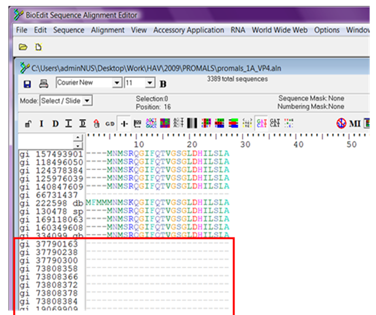
The degree of conservation can also be given as a number for either an individual sequence location or the entire sequence with larger numbers meaning a higher degree of conservation. The amount of conservation of each position is represented by symbols at the bottom of each set of proteins: In the figure, an '*' (asterisk) represents an exact match in all sequences, a ':' (colon) means that a "conserved substitution has occurred" (i.e., the amino acids are not exactly the same, but are of the same general type), and a '.' (period) means that a "semi-conserved substitution has occurred". The canonical representation of an MSA has each protein sequence on a separate line with matching characters aligned in columns and spaces inserted where necessary to improve the alignment – these spaces are called gaps and are represented by dashes. Figure 1 shows a portion of a multiple sequence alignment of DNA methyltransferase (Trdmt1) proteins from 3 different organisms: Human, cow, and mouse. Multiple sequence alignment (MSA) is one of the most widely used methods for assessing sequence conservation and conservaton of protein domains because it allows biologists to analyze the similarities and differences of related proteins at from the individual amino acid level to the sequence level. The MSAVis software, written in Python and using BioPython and OpenGL, can be found at. These visual cues are preattentive and separable so that the relationship between conservation strength and domain membership can be understood.

MSAVis utilizes two visual cues, luminance and hue, to facilitate at-a-glance summary of the conservation of a user-provided protein alignment while enabling multiple comparisons among functional domains. The visualization quickly identifies conserved domains, and allows both macro (sequence-long) and micro (small amino-acid neighborhood) views. The NCBI Conserved Domain Database (CDD) is used for finding conserved domains along the alignment. Input for the algorithm is a multiple sequence alignment in a standard format.

We present MSAVis, a new approach combining luminance and hue for simultaneous visualization of conserved motifs and sequence alignment. By fusing this metadata visually, biologists can analyze sequence conservation and functional motifs simultaneously and efficiently. This insight can be augmented by joint display of conserved domains along the sequences. Multiple alignment of protein sequences can provide insight into sequence conservation across many species and thus allow identification of those sections of the sequence most critical to protein function.


 0 kommentar(er)
